This species has been observed on Reunion and Mayotte Islands
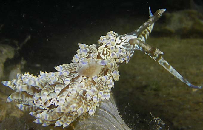
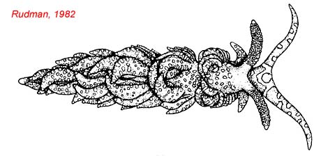
Species characteristics : Background colour is brown or brownish-green with a pattern of light, sometimes whitish patches over cerata and body. Also a distinctive pattern of bands on the head The background colour is caused by the contents of fine branches of the digestive gland which ramify throughout the body wall There is broad yellowish band just below the tip of the cerata. In some specimen there is a blue to purplish band below the yellow band. White cap tip the cerata The rhinophores are papillate except for a narrow line down the anterior midline which devoid of papillae. |
|
|
| Showing species characteristics... | Photo Stephane Ciccione Réunion, "Kelonia center", at Saint Leu, 31 March 2009, size : 60 mm |
|
Remarks :
Identification confirmed by Nathalie Yonow
- Baeolidia major, Eliot, 1903
- Baeolidia major amakusana, Baba, 1937
- Berghia major (Eliot, 1903)
- Spurilla major (Eliot, 1903)
Distrinbution : Tropical iIndo West Pacific Distribution : Australie and Red seaa Cerata : There is a broad yellowish band just below the tip of the cerata . In some specimen there is a blue to purplish band below the yellow band.
The cerata are leaf-like and flattenedCerata : Below the tip is a thin, bright orange band, and a broad blue band which completely rings the cerata . The lower edge of this blue band is irregular, the blue gradually diffusing into the brown.
The cerata are leaf-like and flattened but somewhat swollenHead : There is (sometimes) a distinctive large patch, dark green brown , at the anteriro midline, surrounded by a light, whitish ring.
Head : There is (sometimes) a distinctive large patch, blue , at the anteriro midline, surrounded by a light, whitish ring.
Rhinophores : They are papillate exept for a narrow line down the anterior midline which devoid of papillae.
Rhinophores : They are large and papillate, bearing papillae both anteriorly and posteriorly . They are darker.
Head : There are two bumps (divided swellings ) at the front of the head
Head : No bumps (divided swellings) at the front of the head
Bibliographic data :

References :
Bill Rudman Seaslug site : Sea Slug Forum : Spurilla australis or major
Nudipixel Baeolidia major
Publications :
Other photos of Baeolidia moebii :
.jpg) |
Paul Giannasi Mayotte, cage aquacole de Longoni, 12 m, 12 December 2016, size : 35-40 mm
|
Philibert Bidgrain "Kelonia center", at Saint Leu, 1 April 2009, size : 60 mm (specimen found by Stephane Ciccionne) |
Background colour is brown or brownish-green with a pattern of light, sometimes whitish patches over cerata and body. Also a distinctive pattern of bands on the head |
 |
|
 |
Philibert Bidgrain "Kelonia center", at Saint Leu, 23 March 2009, size : 60 mm (specimen found by Stephane Ciccionne) The anterior edge of te foot has a deep lateral groove (b) forming a distinct upper veil. This veil is split by a median notch (c) forming two bumps (divided swellings). The anterior corners (a) of the foot extend into angular processes. The rhinophores are pale watery brown with darker brown tubercles (d). On close examination one can see brown ducts (e) going up to the stalk and branching into the papillae.
|

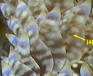
Philibert Bidgrain "Kelonia center", at Saint Leu, 23 March 2009, size : 60 mm (specimen found by Stephane Ciccionne)
In this photo, we can clearly see, below the white tip, a yellowish band (a), We can probably see through the essentially translucent body wall the mass egg (b)  |
 |
 |
Philibert Bidgrain "Kelonia center", at Saint Leu, 23 March and 1 April 2009, size : 60 mm (specimen found by Stephane Ciccionne) We can see through the essentially translucent body wall in the cerata (a) and the foot sole (b) a brown coloration. The brown network pattern are a network of ducts in the skin which contain brown zooxanthellae.
|
Kate Ferrer Reunion, Saint Pierre lagoon, less 1 m, 11 April 2012, size : 20 mm
There is broad yellowish band just below the tip of the cerata and white cap tip the cerata In this specimen there isn't a blue to purplish band below the yellow band.
|
 |
More photos from Indian Ocean
Reunion, Spurilla major, at Etang salé, by Seb Vasquez