This species has been observed on Mayotte Island
The body of the living animal, including rhinophores, oral tentacles, cerata and foot are predominantly a bluish gray color |

|
|
| Showing species characteristics... | Photo Norbert Verneau |
|
See more about : Sightening and mating periods
See more about : Phyllodesmium phylogeny by Moore & Gosliner, 2009
Remarks :
Identification confirmed by Nathalie Yonow
Bibliographic data :
- With arches (1, 2 and 3) consisting of two rows of cerata forming in the anterior ceratal groups and rows (4, 5 and 6) in the most posterior groups.
- The precardiac cerata (1) are grouped into one arch on each side of the body containing approximately 15 cerata. On the arms of the precardiac arches there are double rows of cerata.
- The genital aperture is located between the arms of the precardiac arch on the right side of the animal, and there are separate openings for the male and female genital systems.
- The renal opening is situated between the precardiac arch and the first postcardiac arch on the right side.
- The postcardiac cerata are grouped on both sides into arches containing approximately 13 cerata for the first two arches. These first two postcardiac arches (2 and 3) also contain double rows.
- The anal papilla is located within the first postcardiac arch (2) on the right side.
- The last three ceratal groups (4, 5 and 6) are curved rows containing 4, 3, and 3 cerata respectively.

P. hyalinum |
P. tuberculatum |
P. lembehensis |
P. lizardensis |
|||
| Cerata shape |
Spoon-like, flattened, broadening in upper half. Circular in cross section at the base |
Slightly flattened |
Quadrangular in cross section along the whole length |
Large, thick, and somewhat dorso-ventrally flattened |
Dorso-ventrally flattened with the basal half of each cerata ovate in cross-section |
Dorso-ventrally flattened whereas circular in cross section at the base |
| Cerata tubercles |
Covered with large spiny tubercles |
Low often barely raised, conical tubercles Smooth at the base and only slightly nodulose in the apical part |
Low often barely raised, conical tubercles They are mainly arranged in rows, especially along the edges |
Notably spherical and highly raised They appear with a concentration near the apices |
Tubercles that congregate predominantly on the margins of the upper part of the cerata, appearing almost like rounded serrations
|
Rounded pustules along each edge and a rounded ridge up the central midline. Smooth at the base |
| Anal papilla |
Dorso-laterally of the second ceratal arch
|
Dorsally of the second ceratal row
|
Inside the second ceratal arch |
Inside the second ceratal arch |
Dorsally in the interhepatic space between the first and second ceratal arch |
Inside the second ceratal arch
|
| Foot anterior corner |
Anterior foot corners angular
|
The anterior foot corners only form slightly rounded extensions |
Developed into tentacular processes
|
Broad with moderately tentacular foot corners |
The anterior foot corners only form slightly rounded extensions |
|
| Digestive gland |
Secondary branches |
Secondary branches |
Primary primary |
Secondary, and possibly tertiary, branches |
Primary primary |
Secondary branches
|
References :
Phyllodesmium sp. 2 Gosliner Behrens & Valdés, 2008: 385
Publications :
Other photos of Phyllodesmium cf tuberculatum :
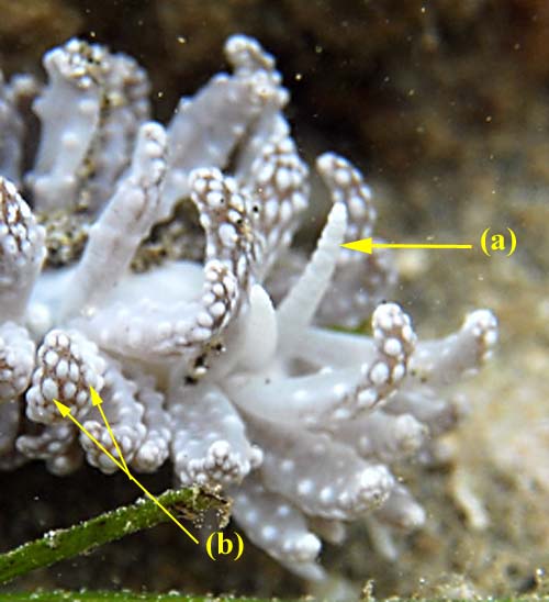 |
Photo Norbert Verneau Mayotte, Petite Terre, Lagon de Badamiers, 1 m, 28 October 2007, size : 20 mm The rhinophores are smooth, with a somewhat wrinkled appearance (a) The tubercles (b) appear on the cerata with a concentration near the apices P. tuberculatum is darker in color than P. hyalinum. So another argument for these identification... |
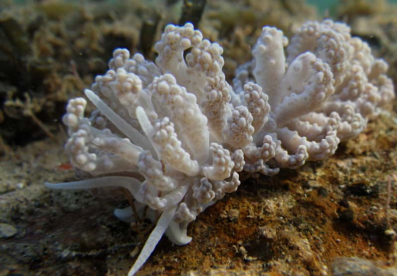
Philibert Bidgrain Mayotte, Petite Terre, déversoir de Dzaoudzi, less 1 m, 17 Jully 2013, size : 25 mm
|
 |
More photos from Indian Ocean
See more about : Phyllodesmium phylogeny by Moore & Gosliner, 2009
If you have taken a photo of this species in Reunion, Mauritius or Mayotte Islands, please Contact us...