This species has been observed on Reunion Mayotte, Madagascar and Seychelles Islands
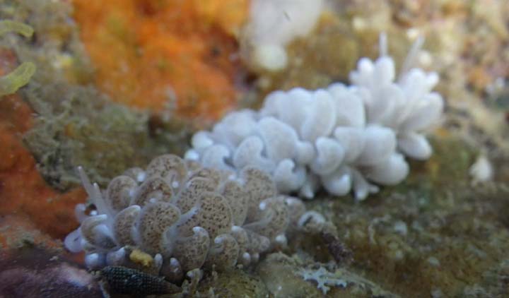
The body wall is a dull translucent white but embedded in it are countless microscopic brownish zooxanthellae which give it and the dorsal side of the cerata a brownish tinge. The purplish tinge to the skin, the yellow tipped rhinophores, oral tentacles and cerata, and yellow border to the foot are all characteristic of that species. The cerata are large, flattened, and partially coiled cerata, with a short cylindrical base and a bluntly pointed tip. |

|
|
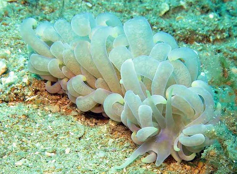
| Showing species characteristics... | Photo Sully Bachel |
|
See more about : Sightening and mating periods
See more about : Phyllodesmium phylogeny by Moore & Gosliner, 2009
See more about : Phyllodesmium ceratal arrangement by Rudman, 1991
See more about : Ceratal structure in Phyllodesmium by Rudman, 1991
Remarks :
Identification confirmed by Nathalie Yonow
- No other name
| Bibliographic data :
|
 |
- These chambers contains clusters of zooxanthellae in those parts of the ceratal wall that are usually unshaded
- Thus zooxanthellae are found on the dorsal surface of large uppermost cerata and on the dorsal and ventral surfaces of the smaller outer cerata
- In the large cerata crowded midway down each row, zooxanthellae are only found in the outer half of the dorsal surface where they are exposed to light
- The concentrations of zooxanthellae appear as brown patches or irregular rings in the living animal
- The zooxanthellae are contained within the cells of the digestive gland
- Zooxanthellae are also found in fine ducts of the digestive gland in the body wall and foot
References :
Bill Rudman Seaslug site : Sea Slug Forum : Phyllodesmium magnum
Nudipixel Phyllodesmium magnum
Publications :
Other photos of Phyllodesmium magnum :
 |
Philibert Bidgrain Mayotte, déversoir de Dzaoudzi, less 1 m, 17 March 2013, size : 20 mm Several specimens observed during the same night.... Mating between two color form of P. magnum
|
Quentin Lognoné Mayotte, Sakouli, less 1 m, 25 March 2017, during the night
|
 |
 |
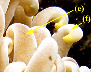
Photo Sully Bachel Reunion, Route en corniche, La Possession, 7 m, 5 February 2010, size : 100 mm The purplish tinge to the skin, the yellow tipped rhinophores (a), oral tentacles (b) and cerata (c), and yellow border (d) to the foot are all characteristic of that species. The concentrations of zooxanthellae (e) appear as brown patches or irregular rings in the living animal The cerata sit slightly enrolled (f)
|
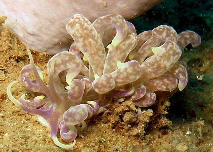
Maurice jay Reunion, Saint Paul The body colour varies depending on the angle from which it is viewed or on which light strikes it. Through some refractive light effect, the body and ceratal wall appear a beautiful bluish lilac (a) possibly from the interaction and reflection of light by the zooxanthellae White patches (b) on the cerata are often present in this species. |
 |
 |
Philippe Bourjon Réunion, Hermitage lagoon, less 1 m,, 28 October 2015, size : 25 mm
|
Alain-Benoit Rassat Madagascar, Nosy Bé, Olaf, 22 m, 25 November 2015, size : 50 mm
|
 |
 |
Alain-Benoît rassat Madagascar, Nosy Bé, size : 120 mm
|
Philippe Bourjon La Réunion , 25 October 2017 |
A trailing behavior ???
|
 |
|
 |
Christophe Mason Parker Seychelles, Baie ternay, Mahé, 12 December 2010.
|
More photos from Indian Ocean
See more about : Phyllodesmium phylogeny by Moore & Gosliner, 2009
See more about : Phyllodesmium ceratal arrangement by Rudman, 1991
See more about : Ceratal structure in Phyllodesmium by Rudman, 1991