This species has been observed on Reunion Island
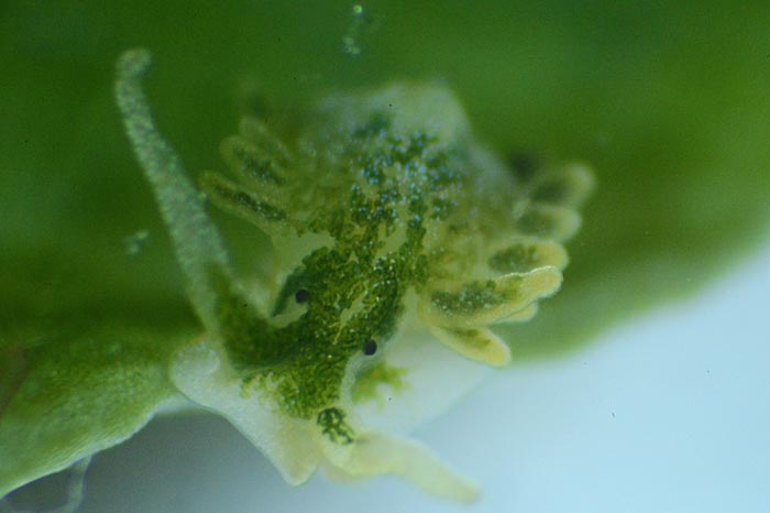
Overall body and head are green with black eyes hardly visible Long green rhinophores with white tips Cerata darker green, darkest at apical end with subapical white blotches forming an incomplete ring. Found in or around in the syncytial algal tubes Boergesenia forbesii |

|
|
| Showing species characteristics... | Photo Philibert Bidgrain |
|
Remarks :
Identification confirmed by Kathe Jensen
- No other name
Bibliographic data :
- Ercolania kencolesi crawled on top of the first third of one algal tube, sat there for about three to four minutes with the ventral part of the head firmly attached to the alga. In these minutes, the slug pierced the algal cell wall and then started to push the head into the alga.
- After seven minutes the slug had penetrated completely into the tube
- It crawled up and down while slurping the cell sap. The mouth opened regularly for this action.
- After 45 minutes, 2/3 of the whole sac was sucked out by the slug
- After 80 minutes it stopped searching and was crawling rather lazily. There are always small patches of chloroplasts which were still recognizable
- After 18 hours, even those patches were gone
- Two days after the intrusion, an egg clutch attached to the algal cell wall was observed
- Then, the slugs left the algal sac and intruded into another algal tube.
- The egg mass is a cylindrical tube (up to 1 mm in diameter) coiled anticlockwise into planar spirals of two and a half whorls
- It is attached to the inner wall of Boergesenia forbesii.
- Usually one animal laid two egg masses inside the same algal sac.
- Egg masses measure 45 mm in diameter and contain around 500 egg capsules, each with one white egg (size about 100 µm) inside. Capsules are rather spherical and measure around 215 µm
References :
Bill Rudman Seaslug site : Sea Slug Forum : Ercolania kencolesi
Publications :
Other photos of Ercolania kencolesi :
 |
Jean-Pascal Quod Réunion, Trois bassins, January 2017, size : 3 mm
|
|
Philibert Bidgrain Reunion, La Saline lagoon, Saint Gilles, less 1 m, 17 February 2010, size: 6 mm After 80 minutes it stopped searching and was crawling rather lazily. There are always small patches of chloroplasts (a) which were still recognizable  |
 Found in or around in the syncytial algal tubes Boergesenia forbesii |
 |
PHilibert Bidgrain Reunion, La Saline lagoon, Saint Gilles, less 1 m, 17 February 2010, size: 6 mm View from the dorsum : Overall colour of body green; under higher magnification, green disintegrating to green dots (a) representing terminal parts of numerous tiny digestive glandular branches. |
View from the sole of the foot : Overall colour of body green; under higher magnification, green disintegrating to green dots (a) representing terminal parts of numerous tiny digestive glandular branches. Anterior margin of foot (b) light green to whitish Cerata darker green, darkest at apical end (c) with subapical white blotches (d) forming an incomplete ring. Long green rhinophores with white tips (e) |
 |
 |
Philibert Bidgrain The slugs left the algal sac Reunion, La Saline lagoon, Saint Gilles, less 1 m, 19 February 2010, size: 9 mm Central notum (a) free of any cerata Cerata darker green, darkest at apical end (b) with subapical white blotches forming an incomplete ring (c). More patches especially in dorsal areas of cerata. Black eyes (d) hardly visible |
Nathalie Rodrigues Reunion, Etang salé on the rocky coast, less 1 m, December 2014
A search of food... the syncytial algal tubes Boergesenia forbesii |
 |
More photos from Indian Ocean
If you have taken a photo of this species in Reunion Island, please Contact us...